History and Development of Microscopes
- Simple microscopes (magnifying glasses) were used in the 13th century.
- Compound microscopes, combining objective lens and eyepiece, appeared in Europe around 1620.
- The inventor of the compound microscope is unknown, with claims made by various individuals.
- Galileo Galilei built his own improved version of the compound microscope after seeing one exhibited by Cornelis Drebbel.
- Giovanni Faber coined the name 'microscope' for the compound microscope.
- The first detailed account of microscopic anatomy appeared in 1644.
- Naturalists in Italy, the Netherlands, and England began using microscopes in the 1660s and 1670s.
- Marcello Malpighi analyzed biological structures, starting with the lungs.
- Robert Hooke's 'Micrographia' had a significant impact due to its illustrations.
- Antonie van Leeuwenhoek achieved up to 300 times magnification using a single lens microscope and made important discoveries.
- In the early 20th century, the electron microscope was developed, using electrons instead of light.
- Ernst Ruska and Max Knoll developed the first prototype transmission electron microscope in 1931.
- The transmission electron microscope uses electromagnets in place of glass lenses.
- The scanning electron microscope was developed in 1935 by Max Knoll.
- Transmission electron microscopes became popular after World War II, and the first commercial scanning electron microscope was developed in 1965.
- Gerd Binnig and Heinrich Rohrer developed a scanning probe microscope at IBM in Zurich, Switzerland.
- In 1986, Binnig and Rohrer received the Nobel Prize in Physics for their invention.
- Marvin Minsky patented the principle of confocal microscopy in 1957.
- Super-resolution microscopy approaches the resolution of electron microscopes.
- STED microscopy was developed by Stefan Hell, Eric Betzig, and William Moerner.
- Technological advances in X-ray lens optics made X-ray microscopes viable in the early 1970s.
Types of Microscopes
- Microscopes can be grouped based on the method they use to interact with a sample and produce images.
- The optical microscope, which uses lenses to refract visible light, is the most common type.
- Fluorescence microscopes, electron microscopes, and scanning probe microscopes are other major types.
- Electron microscopes use electrons instead of light and offer higher resolution.
- Scanning probe microscopes can produce images of small organelles without the need for reagents.
- Atomic force microscopes (AFMs), near-field scanning optical microscopes (NSOM or SNOM), and scanning tunneling microscopes (STMs) are types of scanning probe microscopes.
- Scanning acoustic microscopes use sound waves to measure variations in acoustic impedance.
- Quantum microscopes provide unparalleled precision.
- Mobile app microscopes can be used as optical microscopes when the flashlight is activated.
- Scanning electron microscopes (SEMs) can be used to view the surface of samples.
Applications of Microscopy
- Microscopes are essential tools for studying cells, tissues, and microorganisms in biological research.
- Microscopy helps analyze the structure and properties of materials at the microscopic level in material science.
- Microscopes enable the visualization and manipulation of nanoscale structures in nanotechnology.
- Electron microscopy is used to identify and study viruses in virus detection.
- Microscopes play a crucial role in diagnosing diseases and analyzing tissue samples in medical diagnostics.
Advances in Microscopy
- Aberration-corrected electron microscopy corrects aberrations to achieve higher resolution and image quality.
- Photon-sparse microscopy uses infrared illumination to improve imaging in low-light conditions.
- Scanning probe microscopy allows for atomic-scale imaging and manipulation of surfaces.
- Quantum microscopy uses quantum techniques to study biological processes in living organisms.
- Advances in X-ray and neutron optics enhance the capabilities of X-ray and neutron microscopy.
Resources for Microscopy
- Wikimedia Commons provides a collection of microscope-related media.
- Nature Publishing offers milestones and advancements in light microscopy.
- Nikon MicroscopyU provides tutorials and resources for microscopy.
- Molecular Expressions explores the world of optics and microscopy with educational content.
- Science Daily features articles and news related to microscopy and its applications.
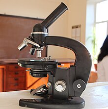
A microscope (from Ancient Greek μικρός (mikrós) 'small', and σκοπέω (skopéō) 'to look (at); examine, inspect') is a laboratory instrument used to examine objects that are too small to be seen by the naked eye. Microscopy is the science of investigating small objects and structures using a microscope. Microscopic means being invisible to the eye unless aided by a microscope.
 | |
| Uses | Small sample observation |
|---|---|
| Notable experiments | Discovery of cells |
| Related items | Optical microscope Electron microscope |
There are many types of microscopes, and they may be grouped in different ways. One way is to describe the method an instrument uses to interact with a sample and produce images, either by sending a beam of light or electrons through a sample in its optical path, by detecting photon emissions from a sample, or by scanning across and a short distance from the surface of a sample using a probe. The most common microscope (and the first to be invented) is the optical microscope, which uses lenses to refract visible light that passed through a thinly sectioned sample to produce an observable image. Other major types of microscopes are the fluorescence microscope, electron microscope (both the transmission electron microscope and the scanning electron microscope) and various types of scanning probe microscopes.
From New Latin microscopium, from Ancient Greek μικρός (mikrós, “small”) + σκοπέω (skopéō, “I look at”).