Signs and symptoms
- Loss of cheek projection with increased width of the face
- Loss of sensation in the cheek and upper lip due to infraorbital nerve injury
- Facial bruising
- Periorbital ecchymosis
- Soft tissue gas, swelling, trismus, altered mastication, diplopia, and ophthalmoplegia as indirect features of the injury
Cause
- Usually caused by a direct blow to the malar eminence of the cheek during assault
- Zygomatic arch fractures at its weakest point, 1.5cm behind the zygomaticotemporal suture
- Zygomas have attachments to the cranium and maxilla, forming the zygomaticomaxillary complex
- Zygomaticomaxillary and zygomaticotemporal sutures are present in the upper and transverse maxillary bone
- Zygomaticomaxillary and frontozygomatic sutures are present in the lateral and vertical maxillary bone
Treatment
- Non-displaced or minimally displaced fractures can be treated conservatively
- Open reduction and internal fixation is reserved for severely angulated or comminuted fractures
- Fixation aims to restore the normal appearance of the face
- Attention is given to the position of the malar eminence and reduction of orbital volume
- Failure to correct the fracture can result in rotational deformity and increased volume of the orbit
Prognosis
- Prognosis of tripod fractures is generally good
- Persistent post-surgical facial asymmetry may occur in some cases
- Further treatment may be required to address facial asymmetry
References
- Fraioli, RE; Branstetter BF, 4th; Deleyiannis, FW (February 2008). Facial fractures: beyond Le Fort.
- Winegar, BA; Murillo, H; Tantiwongkosi, B (2013). Spectrum of critical imaging findings in complex facial skeletal trauma.
- Buchanan, EP; Hopper, RA; Suver, DW; Hayes, AG; Gruss, JS; Birgfeld, CB (December 2012). Zygomaticomaxillary complex fractures and their association with naso-orbito-ethmoid fractures: a 5-year review.
- Swanson, E; Vercler, C; Yaremchuk, MJ; Gordon, CR (May 2012). Modified Gillies approach for zygomatic arch fracture reduction in the setting of bicoronal exposure.
- Linnau, KF; Stanley RB, Jr; Hallam, DK; Gross, JA; Mann, FA (October 2003). Imaging of high-energy midfacial trauma: what the surgeon needs to know.
The zygomaticomaxillary complex fracture, also known as a quadripod fracture, quadramalar fracture, and formerly referred to as a tripod fracture or trimalar fracture, has four components, three of which are directly related to connections between the zygoma and the face, and the fourth being the orbital floor. Its specific locations are the lateral orbital wall (at its superior junction with the zygomaticofrontal suture or its inferior junction with the zygomaticosphenoid suture at the sphenoid greater wing), separation of the maxilla and zygoma at the anterior maxilla (near the zygomaticomaxillary suture), the zygomatic arch, and the orbital floor near the infraorbital canal.
| ZMC complex fracture |
|---|
| Other names | Quadripod fracture |
|---|
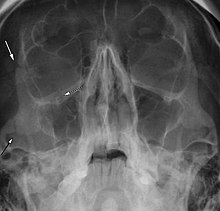 |
| Right zygomaticomaxillary complex fracture with disruption of the lateral orbital wall, orbital floor, zygomatic arch and maxillary sinus. |