Family Friendly & Specialty Dentists in London, UK

Third molars are the most commonly impacted teeth due to their late eruption timing. Maxillary canines rank second in prevalence, accounting for 92.4% of upper jaw impactions. Female patients experience higher impaction rates than males, with notable left-side predominance observed clinically. Various ethnic groups show impaction prevalence ranging from 5.6% to 18.8%. Understanding the specific characteristics and classifications of these impactions provides essential insight into effective treatment approaches.
Although impacted teeth affect approximately 2% of the population, their distribution is far from uniform across dental types.
Upper jaw canines represent a significant concern, accounting for 92.4% of maxillary impacted teeth and 73.2% of all impacted cases overall.
Third molars, however, remain amongst the most commonly impacted teeth due to their late eruption timing.
The prevalence of impacted maxillary canines varies substantially amongst ethnic groups, ranging from 5.6% to 18.8%.
Gender differences also influence impaction rates, with females experiencing higher incidence compared to males.
These variations in prevalence highlight how different teeth present different challenges in the oral cavity.
Understanding these patterns helps clinicians identify populations that may be at greater risk and anticipate potential treatment needs.
Timely intervention through expert dental assessment can help prevent complications such as bite problems and jawbone loss that may result from untreated impacted teeth.
Whilst third molars dominate discussions of tooth impaction due to their frequency, maxillary canines present a distinct clinical challenge that deserves focused attention. Representing a significant proportion of all impacted teeth, impacted canines rank second in prevalence. Clinical observations indicate a notable left-side predominance, with females experiencing higher impaction rates than males. Modern 3D imaging technology can help specialists assess the precise positioning and eruption trajectory of impacted canines before determining the most appropriate treatment approach.
| Characteristic | Clinical Observation |
|---|---|
| Gender Distribution | Higher prevalence in females |
| Lateral Preference | Left-side predominance observed |
| Most Common Type | Type II impactions frequently encountered |
| Secondary Type | Type IV impactions also prevalent |
Contributing factors include insufficient eruption space, jaw size discrepancies, and deep canine germ positioning. Type II impactions feature prominently in clinical presentations, affecting eruption trajectories considerably. Treatment through oral surgery may become necessary when natural eruption fails, requiring careful assessment of underlying anatomical constraints before intervention.
Because impacted teeth present in varied positions and anatomical relationships, standardised classification systems have become essential tools in clinical dentistry.
These frameworks enable practitioners to predict removal challenges and develop appropriate treatment approaches.
The primary classification methods include:
Accurate classification enables clinicians to anticipate removal complications and tailor anaesthesia and extraction strategies accordingly.
The inability of teeth to erupt properly stems from a complex interplay of anatomical, genetic, and developmental factors.
Arch length deficiency represents a primary cause, where insufficient jawbone space prevents teeth from reaching their normal position. Genetic factors significantly influence canine eruption timing and susceptibility to impaction of maxillary canines, with typical eruption occurring around 11-12 years.
Crowding in the dental arch further complicates eruption pathways for commonly impacted teeth, including third molars and maxillary canines. Additional risk factors include cleft palate and delayed tooth development.
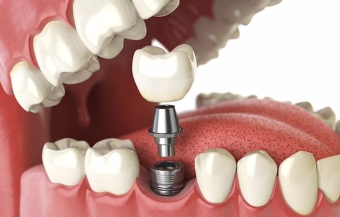
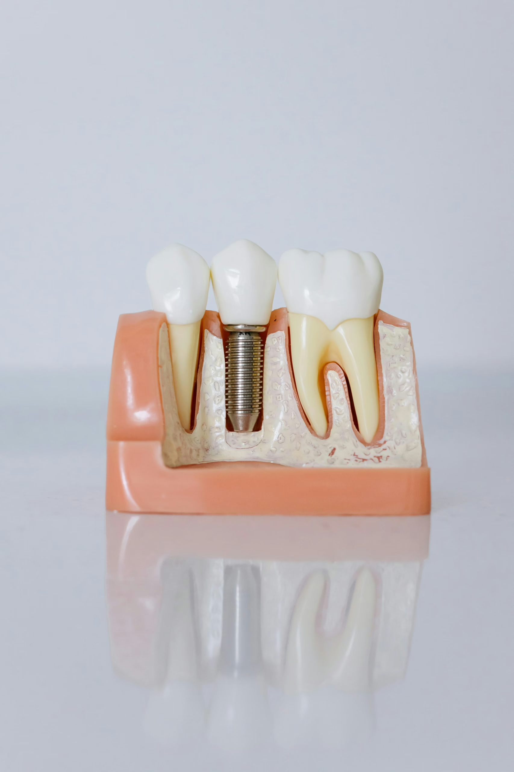
Dental professionals utilise orthopantomograms to assess impaction severity and underlying anatomical constraints, enabling thorough treatment planning that addresses both the impacted tooth and contributing developmental abnormalities. When impacted wisdom teeth cause pain or damage to adjacent structures, surgical extraction procedures may be necessary to prevent further complications and protect overall oral health.
Impacted canines present multifaceted challenges that extend beyond the affected tooth itself, often compromising the health and alignment of surrounding dental structures.
Management requires thorough approaches addressing both immediate complications and long-term outcomes:
Maxillary canines represent the most frequently impacted teeth in the general population, accounting for 73.2% of all impacted dental cases.
These upper canines demonstrate a notable left-side predominance and affect females more frequently than males.
Type II impactions constitute the most common subtype amongst maxillary canine cases, followed by Type IV presentations, making this tooth group clinically significant in orthodontic and surgical practice.
Yes, an impacted tooth may cause swollen lymph nodes in the neck.
When an impacted tooth, such as a third molar or maxillary canine, becomes infected or inflamed, it can trigger pericoronitis or periodontal disease.
The body's immune system responds to this local infection by enlarging nearby lymph nodes as they work to fight the infection.
Persistent or severe lymph node swelling should prompt dental evaluation.
Yes, impacted teeth can cause headaches through multiple mechanisms. The pressure and inflammation from impacted teeth, particularly wisdom teeth and canines, affects surrounding nerves and tissues.
Proximity to nerve pathways creates referred pain, manifesting as headaches or migraines. Infections from partially impacted teeth may also trigger pain radiating to the head and neck.
Dental treatment addressing the impaction may help alleviate these associated headache symptoms.
Type II impaction represents the most common classification of impacted teeth, accounting for 51.6% of all cases.
This classification follows a specific angulation pattern that distinguishes it from other impaction types.
Type IV impaction follows as the second most prevalent, occurring in 28.2% of cases.
These classifications help clinicians understand the spatial relationship and positioning of impacted teeth, which is essential for treatment planning and assessing extraction complexity.
Impacted teeth represent a significant clinical challenge in dentistry, with third molars and maxillary canines accounting for the majority of cases. Understanding the classification systems, risk factors, and potential complications enables practitioners to develop effective management strategies. Early detection through radiographic screening and timely intervention—whether through surgical extraction or orthodontic guidance—may help minimise adverse outcomes and support oral health.