Structure and Course of the Abducens Nerve
- The abducens nucleus is located in the pons, on the floor of the fourth ventricle.
- Axons from the facial nerve loop around the abducens nucleus, creating a slight bulge called the facial colliculus.
- Motor axons leaving the abducens nucleus run ventrally and caudally through the pons.
- The abducens nerve emerges from the brainstem at the junction of the pons and the medulla.
- It runs superior to the medullary pyramid and medial to the facial nerve.
- The nerve enters the subarachnoid space when it emerges from the brainstem.
- It runs upward between the pons and the clivus, and then pierces the dura mater to run between the dura and the skull.
- It makes a sharp turn forward to enter the cavernous sinus and runs alongside the internal carotid artery.
Development and Function of the Abducens Nerve
- The human abducens nerve is derived from the basal plate of the embryonic pons.
- The abducens nerve supplies the lateral rectus muscle of the human eye, responsible for outward gaze.
- It carries axons of type GSE, general somatic efferent.
Clinical Significance of the Abducens Nerve
- Damage to the peripheral part of the abducens nerve can cause double vision and limitation of eye movement.
- Partial damage to the abducens nerve can result in weak or incomplete abduction of the affected eye.
- Fractures of the petrous temporal bone and aneurysms of the intracavernous carotid artery can selectively damage the nerve.
- Infarcts affecting the dorsal pons at the level of the abducens nucleus can also affect the facial nerve, producing an ipsilateral facial palsy together with a lateral rectus palsy.
- Various processes such as tumors, strokes, infections, and neuropathies can damage the abducens nerve.
History and Etymology of the Abducens Nerve
- The Latin name for the sixth cranial nerve is nervus abducens.
- The Terminologia Anatomica recognizes two different English translations: abducent nerve and abducens nerve.
- Abducens is more common in recent literature, while abducent predominates in the older literature.
- The United States National Library of Medicine uses abducens nerve in its Medical Subject Heading (MeSH) vocabulary.
- Grays Anatomy (2005) also prefers abducens nerve.
Abducens Nerve in Other Animals and Related Topics
- The abducens nerve controls the movement of the lateral rectus muscle of the eye.
- In most other mammals, it also innervates the musculus retractor bulbi, which can retract the eye for protection.
- Homologous abducens nerves are found in all vertebrates except lampreys and hagfishes.
- This article uses anatomical terminology.
- References: Grays Anatomy 2008, Chummy S. Sinnatamby (2011), Umansky et al. (1991), Özveren et al. (2007), Halo Orthosis Immobilization – Spine – Orthobullets.
The abducens nerve or abducent nerve, also known as the sixth cranial nerve, cranial nerve VI, or simply CN VI, is a cranial nerve in humans and various other animals that controls the movement of the lateral rectus muscle, one of the extraocular muscles responsible for outward gaze. It is a somatic efferent nerve.
| Abducens nerve | |
|---|---|
 The path of the abducens nerve | |
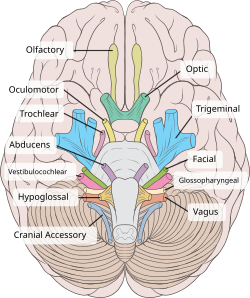
 Inferior view of the human brain, with the cranial nerves labelled. | |
| Details | |
| From | abducens nucleus |
| Innervates | lateral rectus muscle |
| Identifiers | |
| Latin | nervus abducens |
| MeSH | D000010 |
| NeuroNames | 550 |
| TA98 | A14.2.01.098 |
| TA2 | 6283 |
| FMA | 50867 |
| Anatomical terms of neuroanatomy | |
From New Latin nervus abdūcēns (“nerve leading away”).