Family Friendly & Specialty Dentists in London, UK
Presentation and Diagnosis
- Most patients present in the 7th decade of life.
- Females are affected more commonly than males (4:1 ratio).
- The majority of tumors present in the upper lip.
- Few tumors present in the palate or buccal tissue as a slowly enlarging mass.
- Some tumors may show multifocality or multinodularity, which should not be confused with invasion or malignancy.
- Tumors are usually small, with an average size of about 1.6 cm.
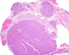
- Histologically, there is a characteristic appearance to the tumor.
- The tumor shows a canalicular pattern with cords and ribbons.
- The connection points between opposing columnar cells within spaces create a 'string of pearls' appearance.
- Small luminal squamous balls or morules are often present, along with a well-developed supporting tissue.
Treatment
- Recurrences are more likely to represent multifocal tumors.
- Conservative surgery is the treatment of choice.
References
- Thompson LD, Bauer JL, Chiosea S, McHugh JB, Seethala RR, Miettinen M, Müller S (Jun 2015). Canalicular adenoma: a clinicopathologic and immunohistochemical analysis of 67 cases with a review of the literature.
- Nelson JF, Jacoway JR (Jun 1973). Monomorphic adenoma (canalicular type). Report of 29 cases.
- Suarez P, Hammond HL, Luna MA, Stimson PG (Aug 1998). Palatal canalicular adenoma: report of 12 cases and review of the literature.
- Penner CR, Thompson LD (Mar 2005). Canalicular adenoma.
- Ferreiro JA (Dec 1994). Immunohistochemical analysis of salivary gland canalicular adenoma.
Other
- Canalicular adenoma must be differentiated from basal cell adenoma, pleomorphic adenoma, adenoid cystic carcinoma, and polymorphous adenocarcinoma.
- Immunohistochemistry studies can be done to confirm the diagnosis.
- Pathologists use pancytokeratin, S100 protein, and SOX10 markers for confirmation.
- The tumor cells show a delicate GFAP reaction around the periphery.
- Small calcifications or microliths may be present in a few cases.
This article may be too technical for most readers to understand. (July 2020) |
Canalicular adenoma is a benign, epithelial salivary gland neoplasm arranged in interconnecting cords of columnar cells. This is a very rare benign neoplasm, that makes up about 1% of all salivary gland tumors, or about 4% of all benign salivary gland tumors.