Structure and Function of Chorda Tympani
- Chorda tympani fibers emerge from the pons of the brainstem as part of the intermediate nerve of the facial nerve.
- The facial nerve exits the cranial cavity through the internal acoustic meatus and enters the facial canal.
- Chorda tympani branches off the facial nerve within the facial canal and enters the middle ear.
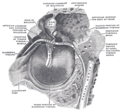
- It runs across the tympanic membrane from posterior to anterior and medial to the neck of the malleus.
- Chorda tympani exits the skull through the petrotympanic fissure and joins the lingual nerve in the infratemporal fossa.
- Chorda tympani carries special sensory fibers providing taste sensation from the anterior two-thirds of the tongue.
- It carries preganglionic parasympathetic fibers to the submandibular ganglion, providing secretomotor innervation to submandibular and sublingual salivary glands.
- Chorda tympani is involved in the dilation of blood vessels in the tongue when stimulated.
- It plays a role in the integration of taste information from facial, glossopharyngeal, and vagus nerves in the nucleus of solitary tract.
- The taste information carried by chorda tympani is mediated by amiloride-sensitive sodium channels.
Taste Sensation and Responses
- Chorda tympani is one of three cranial nerves involved in taste.
- It detects and recognizes sodium chloride most strongly.
- The response to quinine is relatively low in chorda tympani.
- Chorda tympani has varied responses to hydrochloride.
- It is less responsive to sucrose compared to the greater petrosal nerve.
Chorda Tympani Transection
- Chorda tympani shares the nucleus of solitary tract with other cranial nerves.
- When other nerves are cut, chorda tympani takes over the space in the terminal field.
- Transection of chorda tympani at a young age may result in taste buds not growing back fully.
- Bilateral transection of chorda tympani increases preference for sodium chloride.
- Amiloride-sensitive channels responsible for salt recognition are functional in adult rats but not neonatal rats.
Dysfunction of Chorda Tympani
- Injury to the chorda tympani nerve leads to loss or distortion of taste from the anterior two-thirds of the tongue.
- Taste from the posterior one-third of the tongue remains intact.
- Chorda tympani exerts a strong inhibitory influence on other taste nerves and pain fibers in the tongue.
- Damage to chorda tympani disrupts its inhibitory function, leading to less inhibited activity in other nerves.
- Further expansion of this section is needed.
Resources for Studying Chorda Tympani Anatomy
- Wikimedia Commons: Contains media related to Chorda tympani. Provides visual resources for studying the anatomy of Chorda tympani. Can be accessed online. Offers a variety of images and illustrations. Useful for understanding the structure and function of Chorda tympani.
- Anatomy Figure at Human Anatomy Online: Figure 27:03-08 provides information about Chorda tympani. Available on the Human Anatomy Online website. Created by SUNY Downstate Medical Center. Helps in studying the anatomy of Chorda tympani. Offers a visual representation of Chorda tympani.
- Cranial Nerves at Yale School of Medicine: Archived webpage from Yale School of Medicine. Provides information about cranial nerves. Includes details about Chorda tympani. Original page was published on March 3, 2016. Offers a comprehensive overview of cranial nerves.
- MedEd at Loyola Gross Anatomy: MedEd section on Loyola Gross Anatomy website. Contains resources related to Chorda tympani. Focuses on the gross anatomy of Chorda tympani. Offers in-depth information about its structure and function. Useful for studying Chorda tympani in a medical context.
- The Anatomy Lesson by Wesley Norman (Georgetown University): Website by Wesley Norman, affiliated with Georgetown University. Provides information about cranial nerves. Includes a section dedicated to Chorda tympani (CN VII). Offers a photo of Chorda tympani for reference. Useful resource for studying the anatomy of Chorda tympani.
Chorda tympani is a branch of the facial nerve that carries gustatory (taste) sensory innervation from the front of the tongue and parasympathetic (secretomotor) innervation to the submandibular and sublingual salivary glands.
| Chorda tympani | |
|---|---|
 The left tympanic membrane with the malleus and the chorda tympani, viewed from within the tympanic cavity (medial). | |
| Details | |
| From | Facial nerve |
| Innervates | Taste (anterior 2/3 of tongue) Sublingual gland |
| Identifiers | |
| Latin | Nervus chorda tympani |
| MeSH | D002814 |
| TA98 | A14.2.01.084 A14.2.01.118 |
| TA2 | 6292 |
| FMA | 53228 |
| Anatomical terms of neuroanatomy | |
Chorda tympani has a complex course from the brainstem, through the temporal bone and middle ear, into the infratemporal fossa, and ending in the oral cavity.
Borrowed from New Latin chorda tympanī (“cord of the tympanum”).
chorda tympani (plural chordae tympanorum)