Clinical significance and iatrogenic damage
- The marginal mandibular nerve can be injured during neck surgeries, such as submandibular salivary gland excision or neck dissections.
- Lack of accurate knowledge of variations in the nerve's course, branches, and relations contributes to the risk of injury.
- Surgical damage to the nerve can lead to distorted facial expressions, particularly affecting the smile.
- The nerve is located superficially to the facial artery and facial vein, making the artery a useful landmark during surgical procedures.
- Paralysis of the three muscles supplied by the nerve can result from damage, causing an asymmetrical smile due to the lack of contraction of the depressor labii inferioris muscle.
Corrective measures
- Resection of the depressor labii inferioris muscle can be performed to correct the asymmetrical smile caused by paralysis of the marginal mandibular nerve.
- Muscle resection tends to be a successful treatment for this condition.
- Surgical correction of the nerve damage can restore normal facial expressions, including the smile.
- Corrective measures aim to address the lack of contraction in the affected muscles.
- Successful correction of the asymmetrical smile contributes to improved facial aesthetics and function.
Additional images
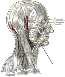
- Lateral head anatomy detail image.
- Neonatal dissection image showing lateral head anatomy detail.
- Images provide visual representations of the marginal mandibular branch of the facial nerve.
- Visual references aid in understanding the anatomical structure and location of the nerve.
- Images can be useful for educational purposes in the study of facial anatomy.
References and external links
- The 20th edition of Gray's Anatomy incorporates text about the marginal mandibular branch of the facial nerve.
- Drake's Gray's Anatomy of Students textbook provides information about the nerve.
- A study published in the Indian Journal of Plastic Surgery explores the anatomical aspects of the nerve.
- The British Journal of Plastic Surgery features a study on the effectiveness of depressor labii inferioris resection as a treatment for nerve paralysis.
- External links provide additional resources for studying the branches of the facial nerve and cranial nerves.
The marginal mandibular branch of the facial nerve arises from the facial nerve (CN VII) in the parotid gland at the parotid plexus. It passes anterior-ward deep to the platysma and depressor anguli oris muscles. It provides motor innervation to muscles of the lower lip and chin:[citation needed] the depressor labii inferioris muscle, depressor anguli oris muscle, and mentalis muscle. It communicates with the mental branch of the inferior alveolar nerve.[citation needed]
| Marginal mandibular branch of the facial nerve | |
|---|---|
 Plan of the facial and intermediate nerves and their communication with other nerves. (Labeled at center bottom, second from bottom, as "Mandibular".) | |
 The nerves of the scalp, face, and side of neck. | |
| Details | |
| From | facial nerve |
| Identifiers | |
| Latin | ramus marginalis mandibularis nervi facialis |
| TA98 | A14.2.01.113 |
| TA2 | 6305 |
| FMA | 53365 |
| Anatomical terms of neuroanatomy | |