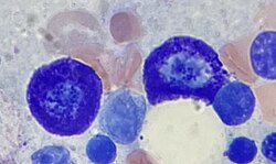
Structure and Distribution of Mast Cells
- Mast cells are granulated cells that contain histamine and heparin.
- They have a round nucleus, unlike basophil granulocytes.
- Mast cells and basophils share a common precursor in bone marrow expressing the CD34 molecule.
- Mast cells settle in different tissue sites, which determines their characteristics.
- Mast cells are present in most tissues, particularly surrounding blood vessels, nerves, and lymphatic vessels.
Function and Mediators of Mast Cells
- Mast cells play a key role in the inflammatory process.
- They can release inflammatory mediators when activated.
- Mast cells can be stimulated by allergens, physical injury, microbial pathogens, and various compounds.
- They express a high-affinity receptor (FcεRI) for the Fc region of IgE.
- Histamine released by mast cells dilates blood vessels and increases permeability.
- Mast cells can release a variety of cytokines and inflammatory mediators.
- Mast cell granules carry bioactive chemicals that can be transferred to adjacent cells.
- Histamine release causes local edema, redness, and itching or pain.
- Mast cells may have a fundamental role in innate immunity.
Mast Cells in the Nervous System
- Mast cells naturally occur in the human brain and interact with the neuroimmune system.
- They are located in structures involved in visceral sensory functions and along the blood-cerebrospinal fluid barrier.
- Mast cells in the brain serve similar functions as in the body, including allergic responses, immunity, and inflammation.
- Mast cells play a role in the gut-brain axis and can be affected by pathogens.
- Mast cells in the nervous system are located near meningeal nociceptors.
Mast Cells in the Gut
- Mucosal mast cells in the gastrointestinal tract are located near sensory nerve fibers.
- Mast cell degranulation releases mediators that activate and sensitize nociceptors.
- Histamine, tryptase, and serotonin are examples of mediators released by mast cells in the gut.
- Mast cells in the gut communicate bidirectionally with sensory nerve fibers.
- Mast cells in the gut play a role in regulating pain and inflammation.
Physiology and Activation of Mast Cells
- FcεR1 is a tetramer composed of one alpha (α) chain, one beta (β) chain, and two gamma (γ) chains.
- IgE binds to the alpha (α) chain of FcεR1, initiating signal transduction through ITAM motifs on the beta (β) and gamma (γ) chains.
- Type 2 helper T cells (Th2) and certain cell types lack the beta (β) chain, relying solely on the gamma (γ) chain for signaling.
- The assembly of the alpha (α) chain with the beta (β) and gamma (γ) chains allows the complex to be exported to the plasma membrane.
- In humans, only the gamma (γ) complex is needed to counterbalance the alpha (α) chain ER retention.
- FcεR1 is a high-affinity IgE receptor found on the surface of mast cells.
- It is a tetramer consisting of one alpha (α) chain, one beta (β) chain, and two identical gamma (γ) chains.
- The alpha (α) chain contains the binding site for IgE and has two domains similar to Ig.
- The beta (β) chain has a single ITAM in the cytoplasmic region, while each gamma (γ) chain also has an ITAM.
- Phosphorylation of the ITAMs by tyrosine initiates the signaling cascade from the receptor.
- Allergen-mediated FcεR1 cross-linking signals resemble the signaling event in antigen binding to lymphocytes.
- Lyn tyrosine kinase is associated with the cytoplasmic end of the FcεR1 beta (β) chain.
- Antigen cross-linking leads to phosphorylation of the ITAMs on the beta (β) and gamma (γ) chains.
- Phosphorylated ITAMs recruit Syk tyrosine kinase, which activates multiple proteins in the signaling cascade.
- Activation of other proteins in the FcεR1-mediated signaling cascade occurs due to antigen-stimulated phosphorylation.
- Linker for activation of T cells (LAT) is an adaptor protein activated by Syk phosphorylation.
- Phospholipase C gamma (PLCγ) becomes phosphorylated when bound to LAT, leading to phosphatidylinositol bisphosphate breakdown.
- Phosphorylation of PLCγ yields inositol trisphosphate (IP3) and diacylglycerol (DAG).
- IP3 elevates calcium levels, while DAG activates protein kinase C (PKC).
- PKC phosphorylates myosin light-chain, facilitating granule movements and fusion with the plasma membrane.
- MRGPRX2 is a G-protein-coupled receptor specific to human mast cells.
- It plays a role in recognizing pathogen-associated molecular patterns (PAMPs) and initiating an antibacterial response.
- MRGPRX2 can bind to competence stimulating peptide (CSP) 1, a quorum sensing molecule produced by Gram-positive bacteria.
- Activation of MRGPRX2 leads to mast cell activation and release of antibacterial mediators.
- MRGPRX2 is a potential therapeutic target and can be activated using the agonist compound 48/80 to control bacterial infection.
Functions and Disorders of Mast Cells
- Mast cells release histamine during anaphylaxis, causing vasodilation and potentially life-threatening shock.
- Mast cells are implicated in autoimmune and inflammatory disorders of the joints, such as rheumatoid arthritis.
- Mastocytosis is a rare disorder characterized by an excessive number of mast cells.
- Mutations in the c-Kit gene are associated with mastocytosis.
- Mast cell tumors, known as mastocyt
Merriam-Webster Online Dictionary
mast cell (
noun)
a granulocyte that occurs especially in connective tissue and has basophilic granules containing substances (as histamine and heparin) which mediate allergic reactions
A mast cell (also known as a mastocyte or a labrocyte) is a resident cell of connective tissue that contains many granules rich in histamine and heparin. Specifically, it is a type of granulocyte derived from the myeloid stem cell that is a part of the immune and neuroimmune systems. Mast cells were discovered by Paul Ehrlich in 1877. Although best known for their role in allergy and anaphylaxis, mast cells play an important protective role as well, being intimately involved in wound healing, angiogenesis, immune tolerance, defense against pathogens, and vascular permeability in brain tumors.
The mast cell is very similar in both appearance and function to the basophil, another type of white blood cell. Although mast cells were once thought to be tissue-resident basophils, it has been shown that the two cells develop from different hematopoietic lineages and thus cannot be the same cells.
English
Etymology
From German Mastzelle f, "feeding cell," coined in 1878 by immunologist Paul Ehrlich.
Noun
mast cell (plural mast cells)
- A resident cell of connective tissue that contains many granules rich in histamine and heparin.
Synonyms
Related terms
...
Read More
