Anatomy and Microanatomy
- The MNTN is located in the brainstem, near the periaqueductal gray and lateral to the cerebral aqueduct.
- It is the only structure in the central nervous system to contain the cell bodies of first-order sensory neurons.
- The mesencephalic nucleus contains no chemical synapses but is electrically coupled.
- Neurons in this nucleus are pseudounipolar and receive proprioceptive afferent information from the mandible.
- They send efferent projections to the trigeminal motor nucleus to mediate monosynaptic jaw jerk reflexes.
Development
- The pseudounipolar neurons in the mesencephalic nucleus are embryologically derived from the neural crest.
- Instead of joining the trigeminal ganglion, the neurons migrate into the brainstem.
- The MNTN is believed to represent a primary sensory ganglion that becomes incorporated into the brainstem during embryologic development.
Function
- The MNTN is involved in reflex proprioception of the periodontium and the muscles of mastication in the jaw.
- Mechanoreceptive nerves in the periodontal ligament sense tooth movement and project to the mesencephalic nucleus.
- Afferent fibers from muscle spindles, the sensory organs of skeletal muscle, are stimulated by the stretch of jaw muscles.
- The mesencephalic nucleus is one of four trigeminal nerve nuclei, involved in conscious facial touch and pain/temperature perception.
- The trigeminal motor nucleus innervates the muscles of mastication and other associated muscles.
Clinical significance
- The mesencephalic nucleus can be tested with the jaw jerk reflex to assess its function.
- Lesions of the trigeminal mesencephalic nucleus can affect feeding due to its role in oral proprioception.
- The mesencephalic nucleus helps prevent excessive biting that may damage the dentition.
Unique features
- The mesencephalic nucleus can be considered functionally as a primary sensory ganglion embedded within the brainstem, making it neuroanatomically unique.
The mesencephalic nucleus of trigeminal nerve is one of the sensory nuclei of the trigeminal nerve (cranial nerve V). It is located in the brainstem. It receives proprioceptive sensory information from the muscles of mastication and other muscles of the head and neck. It is involved in processing information about the position of the jaw/teeth. It is functionally responsible for preventing excessive biting that may damage the dentition, regulating tooth pain perception, and mediating the jaw jerk reflex (by means of projecting to the motor nucleus of the trigeminal nerve).
| Mesencephalic nucleus of trigeminal nerve | |
|---|---|
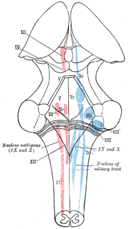
 The cranial nerve nuclei schematically represented; dorsal view. Motor nuclei in red; sensory in blue. (Trigeminal nerve nuclei are at "V".) | |
| Details | |
| Identifiers | |
| Latin | nucleus mesencephalicus nervi trigemini |
| NeuroNames | 558 |
| NeuroLex ID | birnlex_1010 |
| TA98 | A14.1.05.409 |
| TA2 | 5887 |
| FMA | 54568 |
| Anatomical terms of neuroanatomy | |
The axons of the neuron cell bodies of this nucleus provide sensory innervation to target tissues directly, whereas other sensory nuclei of the trigeminal nerve receive their sensory inputs by synapsing with primary sensory neurons in the trigeminal ganglion.