Structure and Origin
- The oculomotor nerve originates from the third nerve nucleus at the level of the superior colliculus in the midbrain.
- The fibers from the third nerve nuclei located laterally on either side of the cerebral aqueduct pass through the red nucleus.
- From the red nucleus, fibers pass via the substantia nigra to emerge from the substance of the brainstem at the oculomotor sulcus.
- The nerve is invested with a sheath of pia mater and enclosed in a prolongation from the arachnoid.
- It passes between the superior cerebellar and posterior cerebral arteries and then pierces the dura mater.
Branches
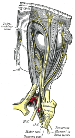
- The superior branch of the oculomotor nerve passes medially over the optic nerve.
- It supplies the superior rectus and levator palpebrae superioris.
- The inferior branch of the oculomotor nerve divides into three branches.
- One passes beneath the optic nerve to the medial rectus.
- Another branch goes to the inferior rectus.
- The third and longest branch runs forward between the inferior recti and lateralis to the inferior oblique.
- A short thick branch is given off to the lower part of the ciliary ganglion.
Nuclei
- The oculomotor nerve arises from the anterior aspect of the mesencephalon (midbrain).
- There are two nuclei for the oculomotor nerve: the oculomotor nucleus and the Edinger-Westphal nucleus.
- The oculomotor nucleus controls the striated muscle in levator palpebrae superioris and other extraocular muscles.
- The Edinger-Westphal nucleus supplies parasympathetic fibers to the eye via the ciliary ganglion, controlling pupil constriction and accommodation.
- Sympathetic postganglionic fibers also join the nerve from the plexus on the internal carotid artery.
Function
- The oculomotor nerve includes axons that innervate skeletal muscles responsible for eye movements.
- It innervates the levator palpebrae superioris, superior rectus, medial rectus, inferior rectus, and inferior oblique muscles.
- The nerve also provides preganglionic parasympathetics to the ciliary ganglion, controlling the constrictor pupillae and ciliary muscles.
- Axons of type GSE (general somatic efferent) and GVE (general visceral efferent) are present in the oculomotor nerve.
- Cranial nerves IV and VI also participate in the control of eye movement.
Additional Information
- Additional images related to the oculomotor nerve.
- Related topics such as anatomical terminology, anisocoria, cranial nerves, and the oculomotor nucleus.
- References and external links for further reading and verification of information.
This article includes a list of general references, but it lacks sufficient corresponding inline citations. (April 2014) |
The oculomotor nerve, also known as the third cranial nerve, cranial nerve III, or simply CN III, is a cranial nerve that enters the orbit through the superior orbital fissure and innervates extraocular muscles that enable most movements of the eye and that raise the eyelid. The nerve also contains fibers that innervate the intrinsic eye muscles that enable pupillary constriction and accommodation (ability to focus on near objects as in reading). The oculomotor nerve is derived from the basal plate of the embryonic midbrain. Cranial nerves IV and VI also participate in control of eye movement.
| Oculomotor nerve | |
|---|---|
 Nerves of the orbit. Seen from above. | |
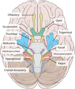
 Inferior view of the human brain, with the cranial nerves labelled. | |
| Details | |
| From | oculomotor nucleus, Edinger-Westphal nucleus |
| To | superior branch, inferior branch |
| Innervates | Superior rectus, inferior rectus, medial rectus, inferior oblique, levator palpebrae superioris, sphincter pupillae (parasympathetics), ciliaris muscle (parasympathetics) |
| Identifiers | |
| Latin | nervus oculomotorius |
| MeSH | D009802 |
| NeuroNames | 488 |
| TA98 | A14.2.01.007 |
| TA2 | 6187 |
| FMA | 50864 |
| Anatomical terms of neuroanatomy | |
oculomotor nerve (plural oculomotor nerves)