Structure and Course
- The trochlear nerve is the fourth cranial nerve.
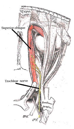
- It innervates the superior oblique muscle of the eye.
- It is the smallest cranial nerve in terms of the number of axons it contains.
- It has the greatest intracranial length.
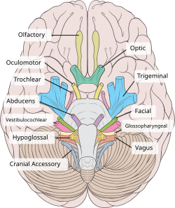
- It exits from the dorsal aspect of the brainstem.
- The trochlear nerve originates from a trochlear nucleus in the medial midbrain.
- It decussates within the midbrain before emerging from the dorsal midbrain.
- It courses on the contralateral side, passing laterally and then anteriorly around the pons.
- It enters the orbit through the superior orbital fissure.
- It innervates the superior oblique muscle.
- The trochlear nerve is derived from the basal plate of the embryonic midbrain.
Clinical Significance and Assessment
- Injury to the trochlear nerve can cause weakness of downward eye movement and vertical diplopia.
- Trochlear nerve palsy also affects torsion or rotation of the eyeball in the plane of the face.
- The characteristic appearance of patients with fourth nerve palsies includes tilting the head to one side and tucking the chin in.
- Causes of trochlear nerve palsy can be both peripheral and central lesions.
- Acute palsy can be caused by head trauma, while chronic palsy is often a congenital defect.
- The trochlear nerve is tested by examining the action of the superior oblique muscle.
- The patient is asked to look down and in, as the superior oblique muscle is most active in this movement.
- Trochlear nerve palsy can result in vertical diplopia, where the affected eye drifts upward relative to the normal eye.
- Patients with trochlear nerve palsy may tilt their head forward or to the opposite side to compensate for torsional diplopia.
- Other causes, such as torticollis, need to be ruled out when diagnosing fourth nerve palsies.
Homologous Trochlear Nerves in Animals
- Trochlear nerves are found in all jawed vertebrates.
- The unique features of the trochlear nerve are seen in the primitive brains of sharks.
- The trochlear nerve has a dorsal exit from the brainstem.
- The trochlear nerve has contralateral innervation.
- Homologous trochlear nerves exist in all jawed vertebrates.
Anatomy of the Trochlear Nerve
- The trochlear nerve has a cavernous portion.
- Traumatic trochlear nerve palsy can occur following minor occipital impact.
- The braincase morphology of the broadnose sevengill shark provides insights into the trochlear nerve.
- Dissection images show the origins of ocular muscles and nerves entering through the superior orbital fissure.
- Deep dissection views reveal the trochlear nerve from a superior perspective.
Additional Resources and Key Points
- Oxford Dictionary on Lexico.com provides a definition of trochlear.
- 'Orbit and accessory visual apparatus: trochlear nerve' is a section in Gray's Anatomy: The Anatomical Basis of Clinical Practice.
- 'Neuroanatomy, Cranial Nerve 4 (Trochlear)' is a publication in StatPearls.
- 'A Textbook of Neuroanatomy' by Maria A. Patestas and Leslie P. Gartner is a comprehensive resource.
- Various research papers and studies provide information on the trochlear nerve.
- NeuroNames provides information on hier-449 related to the trochlear nerve.
- eMedicine offers resources on trochlear nerve palsy.
- Loyola University Medical Education (MedEd) provides educational materials on the trochlear nerve.
- Grossanatomy/h_n/cn/cn1/cn4.htm is a website with information on the trochlear nerve.
- Additional external links related to the trochlear nerve can be found.
- The trochlear nerve is present in all jawed vertebrates.
- Sharks have primitive brains that exhibit unique features of the trochlear nerve.
- The trochlear nerve exits dorsally from the brainstem.
- Contralateral innervation is a characteristic of the trochlear nerve.
- Understanding the anatomy and function of the trochlear nerve is important in various medical contexts.
The trochlear nerve (/ˈtrɒklɪər/), (lit. pulley-like nerve) also known as the fourth cranial nerve, cranial nerve IV, or CN IV, is a cranial nerve that innervates a single muscle - the superior oblique muscle of the eye (which operates through the pulley-like trochlea). Unlike most other cranial nerves, the trochlear nerve is exclusively a motor nerve (somatic efferent nerve).
| Trochlear nerve | |
|---|---|
 The trochlear nerve entering the orbit, seen from above, supplies the superior oblique muscle | |
 The trochlear nerve (CN IV) seen with other cranial nerves. It is the only cranial nerve to emerge from behind the brainstem, and curves around it to reach the front | |
| Details | |
| Innervates | Superior oblique muscle |
| Identifiers | |
| Latin | nervus trochlearis |
| MeSH | D014321 |
| NeuroNames | 466 |
| TA98 | A14.2.01.011 |
| TA2 | 6191 |
| FMA | 50865 |
| Anatomical terms of neuroanatomy | |
The trochlear nerve is unique among the cranial nerves in several respects:
The superior oblique muscle which the trochlear nerve innervates ends in a tendon that passes through a fibrous loop, the trochlea, located anteriorly on the medial aspect of the orbit. Trochlea means “pulley” in Latin; the fourth nerve is thus also named after this structure. The words trochlea and trochlear (/ˈtrɒkliə/, /ˈtrɒkliər/) come from Ancient Greek τροχιλέα trokhiléa, “pulley; block-and-tackle equipment”.
trochlear nerve (plural trochlear nerves)