Family Friendly & Specialty Dentists in London, UK
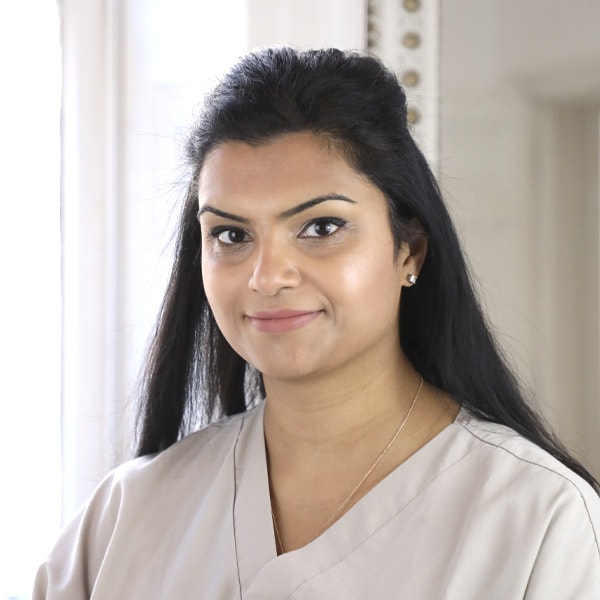
The 3D scan gives a precise map of your root canal anatomy. Dr Neha Patel, specialist in endodontics (GDC No. 150517), uses these scans to identify complex structures that standard X-rays miss.
The scan itself is completed in minutes. A consultation establishes whether this imaging suits your case.
Finding the exact cause of a toothache is often difficult. You may feel severe pain while the source stays hidden from a standard X-ray. We use 3D CBCT imaging to remove this guesswork, seeing the precise anatomy of the tooth and surrounding bone, which matters for endodontics in London. Locating the exact site of an infection or a hidden fracture cuts down the need for exploratory treatment, which matters most for complex cases such as traumatised teeth or cracked tooth syndrome.
Dr Patel uses these scans to build a targeted plan, whether you need root canal retreatment, an apicoectomy, or care for dental trauma, combining them with pulp vitality tests and microscopic examination to confirm the diagnosis before we begin.
Choosing the wrong treatment path is a common worry with complex dental problems. Cone-beam imaging gives us a precise map of the tooth's internal anatomy before we begin, so the diagnosis and the treatment plan are accurate.
We provide a diagnosis-first consultation so we can explain why the scan is clinically necessary and give you a clear breakdown of endodontics costs in London, avoiding unexpected fees.
These scans are essential for finding hidden canals or fractures, and for planning knocked-out tooth treatment. Dr Patel uses these high-resolution images to work through the root system accurately.
Leave your number and we will call you back. Nothing is booked until you say so.
We use the CBCT scan to map a tooth's internal structure before treatment begins. This is often necessary for complex cases where a standard X-ray cannot reveal the full shape of the root canals, and it helps us find hidden canals or fractures that might otherwise cost you the tooth. If you are anxious about the process, we keep the environment calm and supportive. This precision means we can plan pulp capping or follow our root canal aftercare guide more accurately.
Dr Patel assesses your candidacy at the consultation and decides whether this imaging is needed for your specific case.
We begin by capturing high-resolution dental X-rays and 3D images. This lets us map the internal structure of the tooth and surrounding bone, and it is non-invasive. We use these scans to plan complex cases, such as root canal retreatment, where precision matters most.
Our clinicians examine the volumetric data to find hidden canals or fractures, removing the guesswork and showing exactly what is happening beneath the surface. We use this data to decide whether a pulpotomy suits your specific anatomy.
We review the 3D findings at your consultation, using a precise digital model of your tooth to explain the risks and the likely timeline, and to build a plan that protects the natural tooth.
We combine 3D imaging with precision instruments and modern materials to treat infected tooth pulp, and use operating microscopes for a precise diagnosis of tooth fractures in complex cases.
We use bioactive materials to stimulate dentine bridge formation and support pulp healing, and carry out microsurgery to preserve the natural tooth and restore oral health.
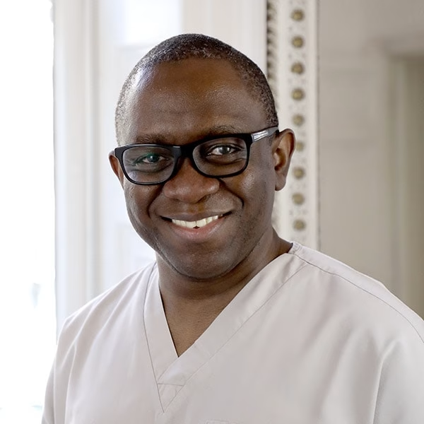
These technologies support root canal retreatment, apicoectomy, cracked tooth treatment, dental trauma care, and treatment for luxation. The practice is led by Dr Ope Sodeinde, GDC-registered specialist periodontist (GDC No. 104071).
We focus on the clinical outcome, with a calm, unhurried manner and honest, no-pressure consultations. This lets us combine 3D scans with a microscopic examination to identify issues that a standard X-ray misses.
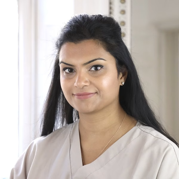
This required root canal treatment (completed by Dr Neha Patel) and then the fitting of a crown, after complex work to reshape the tooth (completed by Dr Ope Sodeinde).
Rather than the extraction, they carefully operated a root canal.
Your consultation establishes the specific requirements of your case and confirms the cost.
Your consultation confirms the timeframe for your specific scan.
The process is non-invasive, and we keep the environment comfortable for patients who feel anxious.
Your consultation determines how long the imaging stays valid for treatment planning, including for a root amputation.
The scan lets us see the exact anatomy of your tooth. This lets Dr Patel plan your treatment with a level of detail a standard X-ray cannot give.
A consultation establishes whether this imaging suits your case. We explain what happens at your root canal consultation and how to book at your first visit.
020 7589 7792
A consultation with Dr Patel establishes whether 3D imaging is necessary for your case. We can also discuss endodontics costs in London, alongside related care: microscopic examination, pulp capping, pulpotomy, root amputation, and knocked-out tooth treatment.
You receive a written plan and an itemised quote from Dr Patel, which is yours to keep and use at any other practice. Arrange your assessment with Dr Patel to find out whether 3D imaging suits your case.
020 7589 7792
Real, verified Google reviews from patients of London Dental Specialists.
Your details are used to arrange your appointment and for nothing else.
111 Regent's Park Road, Primrose Hill, London NW1 8UR
Use our contact page if you would rather ask something than book.