Signs and Symptoms
- Most calcifying odontogenic cysts appear asymptomatic
- Painless, slow-growing mass on the mandible and/or the maxilla
- Swelling in the mouth, both inside the bone and in the gingiva
- Nasal stiffness, epistaxis, and headache when located in the maxilla
- Impacted or displaced teeth and intrabony expansions
Causes and Mechanism/Pathophysiology
- Arises from odontogenic epithelial remnants trapped within the bones or gingival tissues
- Associated with impacted and unerupted teeth
- Presence of ghost cells within the epithelial lining
- Controversial mechanism: coagulative necrosis, enamel protein build-up, or abnormal keratinization
- Formation of large sheets of calcifying material and eosinophilic dentinoid
- Different forms of cystic and neoplastic calcifying odontogenic cysts
Diagnosis
- Radiographic features: unilocular or multilocular radiolucencies with radiopaque deposits
- CT scan reveals internal structures and involvement of neighboring structures
- Histology shows stratified squamous epithelium, stellate reticulum-like cells, and ameloblast-like cells
- Different types of calcifying odontogenic cysts based on special features
- Dental radiographs and CT scans are used for diagnosis
Treatment and Prognosis
- Standard treatment is enucleation and curettage
- Removal of 1 to 2 millimeters layer of bone around the cystic cavity
- Follow-up visits may be required to monitor recurrence
- Rare recurrence after enucleation and curettage
- Favorable prognosis with minimal chance of recurrence
Prevalence and Characteristics
- Only 0.3% of 6,250 oral and maxillofacial lesions diagnosed were calcifying odontogenic cysts
- Most cases found in the mandible
- Age range of affected individuals: 9 to 58 years
- 90% of cases reported no painful symptoms
- 10% of cases reported painful symptoms
- Higher prevalence of calcifying odontogenic cysts in the posterior mandible (55% of cases)
- Other bone diseases commonly found in the posterior mandible, emphasizing the importance of location for diagnosis
- Previous studies found a higher prevalence of calcifying odontogenic cysts in the maxilla and anterior region
- Radiographic and clinical examination crucial for accurate diagnosis
- Histopathological evaluation necessary for confirmation of the diagnosis
Calcifying odontogenic cyst (COC) is a rare developmental lesion that comes from odontogenic epithelium. It is also known as a calcifying cystic odontogenic tumor, which is a proliferation of odontogenic epithelium and scattered nest of ghost cells and calcifications that may form the lining of a cyst, or present as a solid mass.
| Calcifying odontogenic cyst | |
|---|---|
| Other names | Gorlin cyst, calcifying cystic odontogenic tumor |
 | |
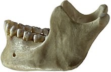
| This condition usually affects the jaw area | |
| Specialty | Dentistry |
It can appear in any location in the oral cavity, but more commonly affects the anterior (front) mandible and maxilla. It is most common in individuals in their 20s to 30s, but can be seen at almost any age, regardless of gender. On dental radiographs, the calcifying odontogenic cyst appears as a unilocular (one circle) radiolucency (dark area). In one-third of cases, an impacted tooth is involved. Histologically, cells that are described as "ghost cells", enlarged eosinophilic epithelial cells without nuclei, are present within the epithelial lining and may undergo calcification.