Anatomy of the Facial Canal:
- The facial canal is the longest bony canal of a nerve in the human body, approximately 3cm long.
- It is located within the middle ear region.
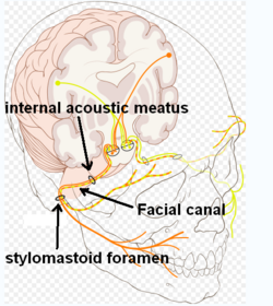
- The canal gives passage to the facial nerve (CN VII) and has a proximal opening at the internal auditory meatus and a distal opening at the stylomastoid foramen.
- While passing through the canal, the facial nerve gives rise to three nerves: the greater petrosal nerve, nerve to stapedius, and the chorda tympani.
Structure of the Facial Canal:
- The horizontal part of the facial canal is divided into two crura: the medial crus (labyrinthine segment) and the lateral crus (tympanic segment).
- The two crura meet at the genu of facial canal, where the geniculate ganglion is situated.
- The lateral crus turns inferior-ward, forming the descending part (mastoid segment) of the canal.
- The descending part of the canal has openings for the nerve to stapedius and the chorda tympani, and it ends at the stylomastoid foramen.
Relations of the Facial Canal:
- The labyrinthine segment of the canal is situated superior to the cochlea.
- The canal traverses the medial wall of the tympanic cavity, located superior to the oval window.
- The prominence of the canal indicates the position of its superior portion.
- The canal curves nearly vertically inferior-ward along the posterior wall.
- The tympanic segment of the canal is closely related to the posterior and medial walls of the tympanic cavity.
Clinical Significance of the Facial Canal:
- Interruptions in the facial canal can lead to variations in the facial nerve.
- Some individuals may have a split facial nerve with 2 or 3 fibers or a poorly formed or absent facial nerve on one side.
History of the Facial Canal:
- The facial canal was first described by Gabriele Falloppio and is sometimes referred to as the Fallopian canal.
The facial canal (also known as the Fallopian canal) is a Z-shaped canal in the temporal bone of the skull. It extends between the internal acoustic meatus and stylomastoid foramen. It transmits the facial nerve (CN VII) (after which it is named).
| Facial canal | |
|---|---|
 Route of facial nerve, with facial canal labeled | |
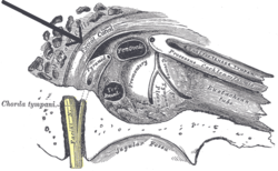
 View of the inner wall of the tympanum. (Facial canal visible in upper left; promontory labeled at center) | |
| Details | |
| System | skeletal |
| Nerve | facial nerve (CN VII) |
| Identifiers | |
| Latin | canalis nervi facialis, canalis facialis |
| TA98 | A02.1.06.009 |
| TA2 | 688 |
| FMA | 54952 |
| Anatomical terminology | |