Gingival Retraction and Gingival Recession
- Gingival retraction is the lateral movement of the gingival margin away from the tooth surface.
- Gingival recession is the spontaneous or non-intentional presentation of gingival retraction.
- Gingival retraction can be performed intentionally using mechanical, chemical, or electrical means for dental surgery procedures.
- Gingival recession may indicate underlying inflammation, pocket formation, or displacement of the marginal gingivae.
- Gingival recession can expose the roots of the teeth.
Gingival Retraction Paste
- Gingival retraction paste provides a dry field and imposes minimal injury on the surrounding periodontium.
- Gingival retraction paste has decreased ability to retract gingival tissues compared to a retraction cord.
- Use of gingival retraction paste is less damaging to the gingival tissues.
- Gingival retraction paste is recommended when minimal retraction is required.
- Gingival retraction paste does not produce haemostasis at the sulcus.
Gingival Retraction Cord
- Gingival retraction cord displaces gingival tissues more effectively compared to retraction paste.
- Gingival retraction cord is recommended when thick periodontium is present.
- Without chemical additions, the retraction cord does not produce haemostasis at the sulcus.
- Gingival retraction cord may cause more damage to the gingival tissues.
- Gingival retraction cord is effective in achieving the desired retraction of gingival tissues.
References
- Mondofacto medical dictionary defines gingival retraction.
- The American Dental Association provides information on the causes and treatment of gingival recession.
- A study published in the Journal of Prosthodontics compares the efficiency of cordless versus cord techniques of gingival retraction.
- The study suggests that both techniques have their advantages and limitations.
- Various dental experts have contributed to the understanding of gingival retraction.
Other
- Capnocytophaga sp. is a type of bacteria associated with periodontal diseases.
- The list of dental experts includes Preston D. Miller, Willoughby D. Miller, Carl E. Misch, John Mankey Riggs, Jay Seibert, Jørgen Slots, Paul Roscoe Stillman, and Dennis P. Tarnow.
- This dentistry article is a stub on Wikipedia.
- The article can be expanded to provide more information on gingival margin.
- The article is categorised under Gingiva and Dentistry stubs.
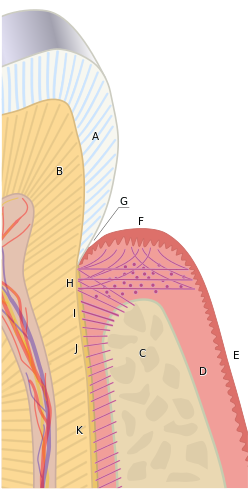
The free gingival margin is the interface between the sulcular epithelium and the epithelium of the oral cavity. This interface exists at the most coronal point of the gingiva, otherwise known as the crest of the marginal gingiva.
| Gingival margin | |
|---|---|
 The gingival margin (F) is the most coronal point of the gingiva, depicted as the zenith of the pink hill in this diagram. To the left lies the sulcular epithelium within the gingival sulcus (G), and to the right lies the oral epithelium (E). | |
| Details | |
| Identifiers | |
| Latin | margo gingivalis |
| TA98 | A05.1.01.109 |
| TA2 | 2791 |
| FMA | 75112 |
| Anatomical terminology | |
Because the short part of gingiva existing above the height of the underlying Alveolar process of maxilla, known as the free gingiva, is not bound down to the periosteum that envelops the bone, it is moveable. However, due to the presence of gingival fibers such as the dentogingival and circular fibers, the free gingiva remains pulled up against the surface of the tooth unless being pushed away by, for example, a periodontal probe or the bristles of a toothbrush.