Structure and Branches of the Glossopharyngeal Nerve
- Glossopharyngeal nerve passes laterally across or below the flocculus
- Leaves the skull through the central part of the jugular foramen
- Has its own sheath of dura mater from the superior and inferior ganglia in jugular foramen
- Inferior ganglion is related to a triangular depression into which the aqueduct of cochlea opens
- Glossopharyngeal nerve is lateral and anterior to the vagus nerve and accessory nerve
- Branches include the tympanic nerve, stylopharyngeal nerve, tonsillar nerve, carotid sinus nerve, branches to the posterior third of the tongue, lingual branches, a communicating branch to the vagus nerve, and contribution to the pharyngeal plexus with the vagus nerve
Overview of Motor Components of the Glossopharyngeal Nerve
- Branchial motor component controls the stylopharyngeus muscle
- Originates from the nucleus ambiguus in the rostral medulla
- Fibers exit the medulla between the olive and the inferior cerebellar peduncle
- Joins other components of CN IX to exit the skull via the jugular foramen
- Innervates the stylopharyngeus muscle to elevate the pharynx during swallowing and speech
- Visceral motor component innervates the ipsilateral parotid gland
- Preganglionic fibers originate in the inferior salivatory nucleus of the rostral medulla
- Fibers exit the brainstem between the medullary olive and the inferior cerebellar peduncle
- Pass through glossopharyngeal ganglia without synapsing
- Lesser petrosal nerve carries the fibers to the otic ganglion and parotid gland
Overview of Sensory Components of the Glossopharyngeal Nerve
- Visceral sensory component innervates the baroreceptors of the carotid sinus and chemoreceptors of the carotid body
- Sensory fibers arise from the carotid sinus and carotid body at the common carotid artery bifurcation
- Join other components of CN IX at the inferior glossopharyngeal ganglion
- Central processes of these neurons enter the skull via the jugular foramen
- Mediates cardiovascular and respiratory reflex responses to changes in blood pressure, CO2, and O2 levels
- Somatic sensory component carries general sensory information from the skin of the external ear, internal surface of the tympanic membrane, walls of the upper pharynx, and posterior one-third of the tongue, anterior surface of the epiglottis, vallecula
- Special sensory component provides taste sensation from the posterior one-third of the tongue
Associated Brainstem Nuclei of the Glossopharyngeal Nerve
- Solitary nucleus: receives taste from the posterior 1/3 of the tongue and information from carotid sinus baroreceptors and carotid body chemoreceptors
- Spinal nucleus of the trigeminal nerve: receives somatic sensory fibers from the internal surface of the tympanic membrane, middle ear, upper part of the pharynx, soft palate, and posterior 1/3 of the tongue
- Nucleus ambiguus: contains lower motor neurons for the stylopharyngeus muscle
- Inferior salivatory nucleus: contains preganglionic parasympathetic neurons to the otic ganglion and then to the parotid gland
Functions and Clinical Significance of the Glossopharyngeal Nerve
- Receives general somatic sensory fibers from the tonsils, pharynx, middle ear, and posterior 1/3 of the tongue
- Receives special visceral sensory fibers (taste) from the posterior 1/3 of the tongue
- Receives visceral sensory fibers from the carotid bodies and carotid sinus
- Supplies parasympathetic fibers to the parotid gland via the otic ganglion
- Supplies motor fibers to the stylopharyngeus muscle and contributes to the pharyngeal plexus
- Damage to the glossopharyngeal nerve can result in loss of taste sensation to the posterior one-third of the tongue and impaired swallowing
- Clinical tests to determine damage include testing the gag reflex, swallowing or coughing, and evaluating speech impediments
- The integrity of the glossopharyngeal nerve may be evaluated by testing general sensation and taste on the posterior third of the tongue
- The gag reflex can also be used to evaluate the glossopharyngeal nerve.
The glossopharyngeal nerve (/ˌɡlɒsoʊfəˈrɪn(d)ʒiəl, -ˌfærənˈdʒiːəl/), also known as the ninth cranial nerve, cranial nerve IX, or simply CN IX, is a cranial nerve that exits the brainstem from the sides of the upper medulla, just anterior (closer to the nose) to the vagus nerve. Being a mixed nerve (sensorimotor), it carries afferent sensory and efferent motor information. The motor division of the glossopharyngeal nerve is derived from the basal plate of the embryonic medulla oblongata, whereas the sensory division originates from the cranial neural crest.
| Glossopharyngeal nerve | |
|---|---|
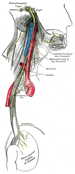
 Plan of the upper portions of the glossopharyngeal, vagus, and accessory nerves. | |
 Course and distribution of the glossopharyngeal, vagus, and accessory nerves. (Label for glossopharyngeal is at upper left.) | |
| Details | |
| To | tympanic nerve |
| Innervates | Motor: stylopharyngeus Sensory: Oropharynx, Eustachian tube, middle ear, posterior third of tongue, carotid sinus, carotid body Special sensory: Taste to posterior third of tongue |
| Identifiers | |
| Latin | nervus glossopharyngeus |
| MeSH | D005930 |
| NeuroNames | 701, 793 |
| NeuroLex ID | birnlex_1274 |
| TA98 | A14.2.01.135 |
| TA2 | 6320 |
| FMA | 50870 |
| Anatomical terms of neuroanatomy | |
glossopharyngeal nerve (plural glossopharyngeal nerves)