Structure and Microanatomy
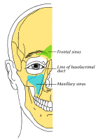
- Maxillary sinus is the largest air sinus in the body with a mean volume of about 10 ml.
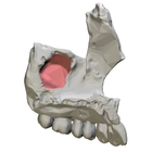
- It is situated within the body of the maxilla and may extend into zygomatic and alveolar processes when large.
- The sinus is pyramid-shaped with the apex at the maxillary zygomatic process.
- The nasal wall presents a large aperture communicating with the nasal cavity, which is reduced in size by various bones.
- The sinus communicates through an opening into the semilunar hiatus.
- The medial wall is primarily composed of cartilage, and the posterior wall has alveolar canals transmitting vessels and nerves.
- The sinus is lined with mucoperiosteum, and the Schneiderian membrane is a bilaminar membrane with ciliated columnar epithelial cells on the internal side.
- The size of the sinuses varies in different skulls and even on the same skull.
Innervation and Relations
- The mucous membranes of the maxillary sinus receive mucomotor parasympathetic nerve fibers from the pterygopalatine ganglion.
- Sensory innervation is provided by the superior alveolar nerves.
- The roof of the sinus is also the floor of the orbit, and posterior to the sinus are the pterygopalatine fossa and infratemporal fossa.
Development and Variation
- The maxillary sinus is the first paranasal sinus to form and rapidly increases in size after puberty.
- The size of the sinus is variable in adults and may extend into zygomatic and alveolar processes.
- The roots of teeth may lie beneath the floor of the sinus or project into the sinus, with the projection more common in advanced age.
- The timing of maxillary sinus growth varies in different people.
Clinical Significance
Subtopic 4.1: Maxillary sinusitis
- Maxillary sinusitis is inflammation of the maxillary sinuses.
- Symptoms include headache, foul-smelling discharge, and systemic signs of infection.
- The skin over the involved sinus can be tender, hot, and reddened.
- Opacification of the sinus on radiographs is due to retained mucus.
- The close anatomical relation to the frontal sinus and maxillary teeth allows for easy spread of infection.
Subtopic 4.2: Oro-antral communication (OAC)
- OAC is an abnormal communication between the maxillary sinus and mouth.
- It is commonly caused by tooth extraction or iatrogenic damage during surgery.
- OAC smaller than 2mm can heal spontaneously, while larger OACs require intervention for closure.
Cancer, Age, and History
- Carcinoma of the maxillary sinus may invade the palate, block the nasolacrimal duct, cause proptosis, spread to the brain, and spread to the lymph nodes.
- With age, the enlarging maxillary sinus may surround the roots of the maxillary posterior teeth and extend into the body of the zygomatic bone.
- Loss of maxillary posterior teeth can further expand the maxillary sinus, thinning the bony floor of the alveolar process.
- Regular dental care becomes crucial to manage age-related changes in the maxillary sinus.
- The maxillary sinus was first discovered and illustrated by Leonardo da Vinci, and Nathaniel Highmore described it in detail in his 1651 treatise.
- Leonardo da Vinci's illustrations and Highmore's treatise played important roles in understanding the maxillary sinus.
English
Noun
maxillary sinus (plural maxillary sinuses)
- (anatomy) A paranasal sinus found in the body of the maxilla.
- Synonym: antrum of Highmore
Translations
Read More

