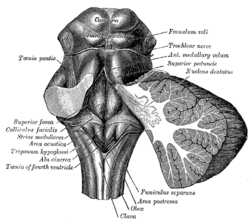
Description and Anatomy of the Fourth Ventricle
- The fourth ventricle is a structure in the brain located in the hindbrain, between the pons and the cerebellum.
- It is filled with cerebrospinal fluid and has a diamond or tetrahedron shape.
- The fourth ventricle is lined with ependymal cells and has a roof, floor, and lateral walls.
- The roof is formed by the cerebellum, the floor by the rhomboid fossa, and the lateral walls contain structures like the vestibular nuclei.
Functions of the Fourth Ventricle
- The fourth ventricle is involved in the regulation of autonomic functions, including respiration and cardiovascular functions.
- It plays a role in the coordination of movements and receives sensory information from the vestibular system.
- The fourth ventricle is important for maintaining balance and posture.
Clinical Importance of the Fourth Ventricle
- Lesions or abnormalities in the fourth ventricle can lead to neurological disorders, such as tumors causing headaches and balance problems.
- Congenital malformations of the fourth ventricle can result in developmental delays.
- Hydrocephalus, the accumulation of cerebrospinal fluid, can affect the fourth ventricle.
- Neurosurgical procedures may be performed to treat conditions affecting the fourth ventricle.
Research and Studying the Fourth Ventricle
- Scientists and researchers study the fourth ventricle to understand its role in neurological functions.
- Imaging techniques like MRI and CT scans are used to visualize the fourth ventricle.
- Animal models are often used for investigating its functions.
- Studying the fourth ventricle can provide insights into the pathophysiology of neurological disorders.
- Research on the fourth ventricle may contribute to the development of new treatment strategies for neurological conditions.
None
Winding around the inferior cerebellar peduncle in the lower part of the fourth ventricle, and crossing the area acustica and the medial eminence are a number of white strands, the medullary striae, which form a portion of the cochlear division of the vestibulocochlear nerve and disappear into the median sulcus. Stria medullaris are axons of arcuate neurons. Courses in the floor of the fourth ventricle. Joins the restiform body to reach the cerebellum.
| Medullary striae of fourth ventricle | |
|---|---|
 Rhomboid fossa (striae medullares labeled at center left) | |
| Details | |
| Identifiers | |
| Latin | striae medullares ventriculi quarti |
| TA98 | A14.1.05.318 A14.1.05.707 |
| TA2 | 6046 |
| FMA | 78484 |
| Anatomical terms of neuroanatomy | |