Structure and Function of the Olfactory Bulb
- The olfactory bulb is located on the inferior side of the brain in humans and is divided into the main olfactory bulb and the accessory olfactory bulb.
- The main olfactory bulb connects to the amygdala and plays a role in odor detection and discrimination.
- Destruction of the olfactory bulb results in ipsilateral anosmia.
- The olfactory bulb has a multi-layered cellular architecture, including the glomerular layer, external plexiform layer, mitral cell layer, internal plexiform layer, and granule cell layer.
- The olfactory bulb enhances the sensitivity of odor detection and filters out background odors to enhance the transmission of select odors.
- The olfactory bulb permits higher brain areas involved in arousal and attention to modify the detection or discrimination of odors.
Temporal Processing and Lateral Inhibition in the Olfactory Bulb
- Processing occurs at each level of the main olfactory bulb, with interneurons in the external plexiform layer exhibiting both excitatory and inhibitory potentials.
- Neural firing in the olfactory bulb varies temporally, and synchronised mitral cell spike trains help discriminate similar odors better.
- Lateral inhibition, mediated by interneurons in the external plexiform layer, aids in odor discrimination by decreasing firing in response to background odors.
- The granule cell layer, which connects mitral cells and interneurons known as granule cells, plays a crucial role in lateral inhibition and differentiated odor responses.
Accessory Olfactory Bulb and Further Processing of Olfactory Information
- The accessory olfactory bulb forms a parallel pathway independent from the main olfactory bulb and receives input from the vomeronasal organ, involved in social and reproductive behaviors.
- Olfactory information from the olfactory bulb is further processed in the amygdala, orbitofrontal cortex (OFC), and hippocampus.
- The amygdala is involved in associative learning between odors and behavioral responses, while the OFC assesses the value and nutritional content of a food reward.
- The hippocampus aids in olfactory memory and learning, including the formation of episodic memory.
Olfactory Coding in Habenula and Depression Models
- Neurons in the hippocampus and habenula fire in response to odor stimuli, and the dorsal habenula shows functional asymmetry with predominant odor responses in the right hemisphere.
- Olfactory bulb removal in rats causes structural changes in the amygdala and hippocampus, leading to behavioral changes similar to depression.
- Decreased neuroplasticity in the hippocampus is observed after olfactory bulb removal, which is associated with behavioral changes characteristic of depression.
Adult Neurogenesis and Role of the Hippocampus and Amygdala
- The olfactory bulb, subventricular zone, and subgranular zone of the hippocampus undergo continuing neurogenesis in adult mammals.
- New neurons in the olfactory bulb may participate in learning processes and are sensitive to olfactory activity.
- The hippocampus and amygdala influence odor perception and associate odors with emotions and rewards.
- The orbitofrontal cortex contributes to appetite regulation and projects to the anterior cingulate cortex.
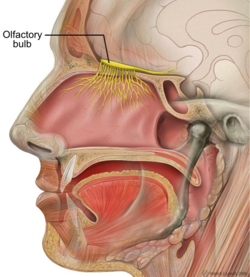
The olfactory bulb (Latin: bulbus olfactorius) is a neural structure of the vertebrate forebrain involved in olfaction, the sense of smell. It sends olfactory information to be further processed in the amygdala, the orbitofrontal cortex (OFC) and the hippocampus where it plays a role in emotion, memory and learning. The bulb is divided into two distinct structures: the main olfactory bulb and the accessory olfactory bulb. The main olfactory bulb connects to the amygdala via the piriform cortex of the primary olfactory cortex and directly projects from the main olfactory bulb to specific amygdala areas. The accessory olfactory bulb resides on the dorsal-posterior region of the main olfactory bulb and forms a parallel pathway. Destruction of the olfactory bulb results in ipsilateral anosmia, while irritative lesions of the uncus can result in olfactory and gustatory hallucinations.
| Olfactory bulb | |
|---|---|
 Human brain seen from below. Vesalius' Fabrica, 1543. Olfactory bulbs and olfactory tracts outlined in red | |
 Sagittal section of human head. | |
| Details | |
| System | Olfactory |
| Identifiers | |
| Latin | bulbus olfactorius |
| MeSH | D009830 |
| NeuroNames | 279 |
| NeuroLex ID | birnlex_1137 |
| TA98 | A14.1.09.429 |
| TA2 | 5538 |
| FMA | 77624 |
| Anatomical terms of neuroanatomy | |

