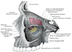
Structure and Boundaries of the Pterygopalatine Fossa
- Anterior boundary: superomedial part of the infratemporal surface of maxilla
- Posterior boundary: root of the pterygoid process and adjoining anterior surface of the greater wing of sphenoid bone
- Medial boundary: perpendicular plate of the palatine bone and its orbital and sphenoidal processes
- Lateral boundary: pterygomaxillary fissure
- Inferior boundary: part of the floor formed by the pyramidal process of the palatine bone
Nerve Supply of the Pterygopalatine Fossa
- The pterygopalatine fossa contains the second division of the trigeminal nerve
- The nerve of the pterygoid canal is a combination of the greater petrosal nerve (preganglionic parasympathetic) and the deep petrosal nerve (postganglionic sympathetic)
- Intraoral injection in this area can lead to block anesthesia of the entire second division of the trigeminal nerve
Related Articles and Terminology
- This article uses anatomical terminology
- Pterygopalatine canal (disambiguation)
- Fossa in the Human Body
Additional Images of the Pterygopalatine Fossa
- Alveolar branches of superior maxillary nerve and pterygopalatine ganglion
- The pterygopalatine ganglion and its branches
- Pterygopalatine fossa in a dog
- Pterygopalatine fossa
References and External Links
- Illustrated Anatomy of the Head and Neck, Fehrenbach and Herring, Elsevier, 2012, page 69
- Osborn, Anne (March 1979). Radiology of the Pterygoid Plates and Pterygopalatine Fossa (PDF). American Journal of Roentgenology. 132 (3): 389–394. doi:10.2214/ajr.132.3.389. PMID106641.
- Ryan, Stephanie (2011). Chapter 1. Anatomy for diagnostic imaging (Thirded.). Elsevier Ltd. p.35. ISBN9780702029714.
- External link: Interactive at Columbia.edu
In human anatomy, the pterygopalatine fossa (sphenopalatine fossa) is a fossa in the skull. A human skull contains two pterygopalatine fossae—one on the left side, and another on the right side. Each fossa is a cone-shaped paired depression deep to the infratemporal fossa and posterior to the maxilla on each side of the skull, located between the pterygoid process and the maxillary tuberosity close to the apex of the orbit. It is the indented area medial to the pterygomaxillary fissure leading into the sphenopalatine foramen. It communicates with the nasal and oral cavities, infratemporal fossa, orbit, pharynx, and middle cranial fossa through eight foramina.
| Pterygopalatine fossa | |
|---|---|
 Left maxillary sinus opened from the exterior. | |
 Human skull with entrance to pterygopalatine fossa marked in red | |
| Details | |
| Identifiers | |
| Latin | fossa pterygopalatina |
| MeSH | D056739 |
| TA98 | A02.1.00.025 |
| TA2 | 429 |
| FMA | 75309 |
| Anatomical terms of bone | |
pterygopalatine fossa (plural pterygopalatine fossae)