Neuroanatomy
- The solitary nucleus is a series of sensory nuclei located in the medulla oblongata.
- It forms a vertical column of grey matter in the brainstem.
- The nucleus receives inputs from cranial nerves VII, IX, and X.
- It is connected by the solitary tract, a bundle of nerve fibers.
- Cell bodies within the nucleus are arranged according to function.
Afferents
- The nucleus receives gustatory (taste) sensation from cranial nerves VII, IX, and X.
- It also receives input from chemoreceptors and mechanoreceptors of the general visceral afferent pathway.
- These receptors are located in various organs such as the carotid body, carotid sinus, aortic bodies, and sinoatrial node.
- Additional minor input comes from the nasal cavity, soft palate, and sinus cavities.
- Organ-specific regions of neuronal architecture are preserved in the solitary nucleus.
Efferents
- The solitary nucleus projects to various regions of the brain, including the hypothalamus, amygdala, and other brainstem nuclei.
- It forms circuits that contribute to autonomic regulation.
- The nucleus sends stimuli related to oral cavity and gastrointestinal tract to the parabrachial area.
- Different subdivisions of the parabrachial area receive gastric and gustatory (taste) processes.
- Some neuronal subpopulations in the solitary nucleus project to the bed nucleus of the stria terminalis.
Function
- Afferents of the solitary nucleus mediate reflexes such as the gag reflex, carotid sinus reflex, aortic reflex, cough reflex, baroreflex, and chemoreceptor reflexes.
- Neurons within the nucleus also transmit signals about the gut wall, lung stretch, and dryness of mucous membranes.
- The nucleus participates in simple autonomic reflexes.
- It regulates motility and secretion within the gastrointestinal system.
- The solitary nucleus is involved in respiratory reflexes.
Additional images
- A section of the medulla oblongata shows the location of the solitary nucleus.
- Primary terminal nuclei of the sensory cranial nerves are represented in a lateral view.
- These images provide visual representations of the anatomical structures.
- They aid in understanding the location and organization of the solitary nucleus.
- The images can be used for educational and reference purposes.
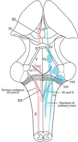
The solitary nucleus (also called nucleus of the solitary tract, nucleus solitarius, or nucleus tractus solitarii (SN or NTS)) is a series of sensory nuclei (clusters of nerve cell bodies) forming a vertical column of grey matter in the medulla oblongata of the brainstem. It receives general visceral and/or special visceral inputs from the facial nerve (CN VII), glossopharyngeal nerve (CN IX) and vagus nerve (CN X); it receives and relays stimuli related to taste and visceral sensation. It sends outputs to various parts of the brain.[where?] Neuron cell bodies of the SN are roughly somatotopically arranged along its length according to function.
| Solitary nucleus | |
|---|---|
 The cranial nerve nuclei schematically represented; dorsal view. Motor nuclei in red; sensory in blue. | |
 Transverse section of medulla oblongata of human embryo. | |
| Details | |
| Identifiers | |
| Latin | Nucleus tractus solitarii medullae oblongatae. |
| MeSH | D017552 |
| NeuroNames | 742 |
| NeuroLex ID | birnlex_1429 |
| TA98 | A14.1.04.230 |
| TA2 | 6008 |
| FMA | 72242 |
| Anatomical terms of neuroanatomy | |