Structure and Origin of the Trigeminal Nerve
- Trigeminal nerve is the fifth cranial nerve responsible for sensation in the face and motor functions such as biting and chewing.
- It has three major branches: ophthalmic, maxillary, and mandibular nerves.
- The ophthalmic and maxillary nerves are purely sensory, while the mandibular nerve supplies motor and sensory functions.
- Autonomic nerve fibers and special sensory fibers (taste) are contained within the trigeminal nerve.
- The trigeminal nerve enters the brainstem at the level of the pons.
- It has a sensory root and a smaller motor root.
- Motor fibers pass through the trigeminal ganglion without synapsing on their way to peripheral muscles.
- The sensory division originates in the cranial neural crest.
- Sensory information is processed in the nucleus of the fifth nerve within the pons.
- The trigeminal ganglion is located within Meckels cave and contains the cell bodies of incoming sensory-nerve fibers.
- It is analogous to the dorsal root ganglia of the spinal cord.
- The ganglion receives input from the ophthalmic, maxillary, and mandibular branches of the trigeminal nerve.
- It is also known as the semilunar ganglion or gasserian ganglion.
- The ganglion is responsible for transmitting sensory information from the face and mouth.
- The ophthalmic, maxillary, and mandibular branches leave the skull through separate foramina.
- The ophthalmic nerve carries sensory information from the scalp, forehead, eyelid, nose, and frontal sinuses.
- The maxillary nerve carries sensory information from the lower eyelid, cheek, upper lip, and maxillary sinuses.
- The mandibular nerve carries sensory information from the lower lip, lower teeth, chin, and external ear.
- The mandibular nerve also carries touch-position and pain-temperature sensations from the mouth.
Function of the Trigeminal Nerve
- The trigeminal nerve provides tactile, proprioceptive, and nociceptive afference to the face and mouth.
- It activates the muscles of mastication and other related muscles.
- The trigeminal nerve carries general somatic afferent fibers (GSA) for skin innervation.
- It also carries special visceral efferent (SVE) axons for muscle innervation.
- The motor component of the mandibular division controls the movement of muscles involved in biting, chewing, and swallowing.
Sensory Pathways and Trigeminal Nuclei
- Sensory information is sent to specific nuclei in the thalamus.
- Thalamic nuclei send information to specific areas in the cerebral cortex.
- Each pathway consists of three bundles of nerve fibers connected in series.
- Secondary neurons in each pathway decussate (cross the spinal cord or brainstem).
- Sensory information is processed and modified at each level in the chain by interneurons and input from other areas of the nervous system.
- The trigeminal nucleus receives sensory information from the face.
- Sensation from parts of the mouth, ear, and meninges is carried by other cranial nerves.
- The trigeminal nucleus extends throughout the brainstem and cervical cord.
- The nucleus is divided into three parts: spinal trigeminal, principal sensory, and mesencephalic nuclei.
- The spinal trigeminal nucleus represents pain-temperature sensation from the face.
- Pain-temperature fibers from peripheral nociceptors are carried in cranial nerves.
- The spinal trigeminal nucleus contains a pain-temperature sensory map of the face and mouth.
- Secondary fibers from the spinal trigeminal nucleus cross the midline and ascend to the contralateral thalamus.
- Pain-temperature fibers are sent to multiple thalamic nuclei.
- The principal nucleus represents touch-pressure sensation from the face.
- Located in the pons, near the entrance for the fifth nerve.
- Receives touch-position information from cranial nerves V, VII, IX, and X.
- Contains a touch-position sensory map of the face and mouth.
- Secondary fibers cross the midline and ascend to the contralateral thalamus.
- The mesencephalic nucleus is a sensory ganglion embedded in the brainstem.
- Contains proprioceptor fibers from the jaw and mechanoreceptor fibers from the teeth.
- Some fibers bypass conscious perception and go directly to the motor nucleus of the trigeminal nerve.
- Plays a role in coordinating biting, chewing, and swallowing.
Pathways to the Thalamus and Cortex
- Sensory input (except smell) is sent to the thalamus and then the cortex.
- Thalamus is anatomically subdivided into nuclei.
- Touch-position information from the body goes to the ventral posterolateral nucleus (VPL) of the thalamus.
- Touch-position information from the face goes to the ventral posteromedial nucleus (VPM) of the thalamus.
- Information is projected to the primary somatosensory cortex (SI) in the parietal lobe.
- Pain-temperature information is sent to the VPL (body) and VPM (face) of the thalamus.
- Pain-temperature information is also sent to other thalamic nuclei and projected onto additional areas of the cerebral cortex.
- Some fibers go to the medial dorsal thalamic nucleus (MD) and project to the anterior cingulate cortex.
- Other fibers go to the ventromedial (VM) nucleus of the thalamus and project to the insular cortex.
- Some fibers go to the intralaminar nucleus (IL) of the thalamus via the reticular formation and project diffusely to all parts of the cerebral cortex.
Clinical Significance and Additional Images
- Trigeminal neuralgia, cluster headache, migraine, and lateral medullary syndrome (Wallenberg syndrome) are clinical conditions associated with the
This article includes a list of general references, but it lacks sufficient corresponding inline citations. (October 2014) |
In neuroanatomy, the trigeminal nerve (lit. triplet nerve), also known as the fifth cranial nerve, cranial nerve V, or simply CN V, is a cranial nerve responsible for sensation in the face and motor functions such as biting and chewing; it is the most complex of the cranial nerves. Its name (trigeminal, from Latin tri- 'three', and -geminus 'twin') derives from each of the two nerves (one on each side of the pons) having three major branches: the ophthalmic nerve (V1), the maxillary nerve (V2), and the mandibular nerve (V3). The ophthalmic and maxillary nerves are purely sensory, whereas the mandibular nerve supplies motor as well as sensory (or "cutaneous") functions. Adding to the complexity of this nerve is that autonomic nerve fibers as well as special sensory fibers (taste) are contained within it.
| Trigeminal nerve | |
|---|---|
 Schematic illustration of the trigeminal nerve and the organs (or structures) it supplies | |
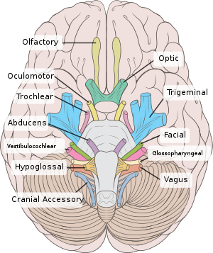
 Inferior view of the human brain, with cranial nerves labelled | |
| Details | |
| To | Ophthalmic nerve Maxillary nerve Mandibular nerve |
| Innervates | Motor: Muscles of mastication, tensor tympani, tensor veli palatini, mylohyoid, anterior belly of the digastric Sensory: Face, mouth, temporomandibular joint |
| Identifiers | |
| Latin | nervus trigeminus |
| MeSH | D014276 |
| NeuroNames | 549 |
| TA98 | A14.2.01.012 |
| TA2 | 6192 |
| FMA | 50866 |
| Anatomical terms of neuroanatomy | |
The motor division of the trigeminal nerve derives from the basal plate of the embryonic pons, and the sensory division originates in the cranial neural crest. Sensory information from the face and body is processed by parallel pathways in the central nervous system.
trigeminal nerve (plural trigeminal nerves)