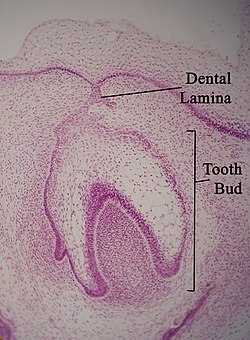
Function and Development of Dental Lamina
- Dental lamina is involved in the development of teeth.
- It is derived from a primary epithelial band that forms when oral epithelium invaginates into the mesenchyme.
- Proliferation on the cranial portion of the dental lamina leads to the formation of deciduous teeth.
- Further proliferation on the leading edge of the lamina results in the development of permanent teeth.
- Permanent molars develop from the general lamina, which is also derived from the dental lamina.
Hyperactivity of Dental Lamina and Supernumerary Teeth
- Hyperactivity of the dental lamina can cause conditions like hyperdontia, where patients have supernumerary teeth.
- Possible reasons for hyperdontia include dichotomy of tooth buds, atavism, Gardner's syndrome, and hyperactivity of the dental lamina.
- The most accepted theory for supernumerary teeth is the hyperactivity of the dental lamina.
- When remnants of the dental lamina fail to resorb, abnormal proliferation can occur, leading to the formation of extra tooth buds and supernumerary teeth.
Dental Lamina and Tooth Eruption
- During the bell stage of tooth development, the dental lamina helps to separate the oral epithelium from the developing tooth.
- Breaking up of the dental lamina can result in the formation of epithelial cell clusters.
- Some of these clusters may persist as epithelial pearls, which can delay tooth eruption by creating cysts on the developing tooth.
- The dental lamina plays a role in the coordination of tooth eruption into the oral cavity.
- Disintegration of the dental lamina allows the tooth to emerge properly.
Dental Lamina and Tooth Development
- The dental lamina is the first evidence of tooth development and begins around the sixth week in utero.
- It is formed when oral ectoderm cells proliferate faster than cells in other areas.
- The dental lamina connects the developing tooth bud to the epithelium of the oral cavity.
- It eventually disintegrates into small clusters of epithelium and is resorbed.
- The dental lamina gives rise to ameloblasts and enamel, while the ectomesenchyme is responsible for the dental papilla and odontoblasts.
Related Concepts
- Diphyodont and polyphyodont are related concepts to dental lamina.
- Diphyodont refers to the development of two sets of teeth, while polyphyodont refers to the continuous replacement of teeth throughout an animal's life.
- Understanding the dental lamina is essential for studying tooth replacement in amniotes.
- Dental lamina regression is a crucial process underlying the development of diphyodont dentitions.
- Various studies and literature reviews have been conducted to explore the etiology and characteristics of supernumerary teeth related to dental lamina hyperactivity.
The dental lamina is a band of epithelial tissue seen in histologic sections of a developing tooth. The dental lamina is first evidence of tooth development and begins (in humans) at the sixth week in utero or three weeks after the rupture of the buccopharyngeal membrane. It is formed when cells of the oral ectoderm proliferate faster than cells of other areas. Best described as an in-growth of oral ectoderm, the dental lamina is frequently distinguished from the vestibular lamina, which develops concurrently. This dividing tissue is surrounded by and, some would argue, stimulated by ectomesenchymal growth. When it is present, the dental lamina connects the developing tooth bud to the epithelium of the oral cavity. Eventually, the dental lamina disintegrates into small clusters of epithelium and is resorbed. In situations when the clusters are not resorbed, (this remnant of the dental lamina is sometimes known as the glands of Serres) eruption cysts are formed over the developing tooth and delay its eruption into the oral cavity. This invagination of ectodermal tissues is the progenitor to the later ameloblasts and enamel while the ectomesenchyme is responsible for the dental papilla and later odontoblasts.
| Dental lamina | |
|---|---|
 Micrograph of a dental lamina and tooth bud. H&E stain. | |
| Details | |
| Identifiers | |
| Latin | lamina dentalis |
| TE | lamina_by_E5.4.1.1.1.0.3 E5.4.1.1.1.0.3 |
| Anatomical terminology | |