Pathogenesis and Clinical Features
- Odontogenesis is the process of tooth development involving interaction between oral epithelium and surrounding mesenchymal tissue.
- Ectopic tooth eruption can occur due to pathological processes or developmental disturbances.
- Accumulation of fluid between the reduced enamel epithelium and enamel or within the layers of enamel organ is key to the formation of dentigerous cysts.
- Rapid transudation of serum across capillary walls can occur when a potentially erupting tooth obstructs venous outflow.
- The exact histogenesis of dentigerous cysts is unknown, but most authors believe it originates from the tooth follicle.
- Dentigerous cysts commonly involve a single tooth, with the mandibular third molar being the most frequently affected.
- Maxillary canines may also be involved, and supernumerary or ectopic teeth can be associated with dentigerous cysts.
- Displacement of involved teeth into ectopic positions, including the maxillary sinus, can occur.
- Maxillary sinus involvement can cause symptoms such as headache, facial pain, purulent nasal discharge, or nasolacrimal obstruction.
- Dentigerous cysts involving premolars are rare, with a reported incidence of 1.44 in every 100 unerupted teeth.
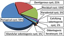
- Dentigerous cysts are the second most prevalent type of odontogenic cysts, after radicular cysts.
- About 70% of dentigerous cysts occur in the mandible.
- The age of presentation ranges from 3 years to 57 years, with a mean age of 22.5 years.
- Males have a higher prevalence than females, with a ratio of 1.8:1.
- Bilateral or multiple dentigerous cysts are rare, except in cases of certain syndromes or drug-induced cysts.
Potential Complications and Treatment
- Dentigerous cysts can become aggressive lesions and cause continuous enlargement.
- Possible complications include expansion of alveolar bone, displacement of teeth, severe root resorption, and expansion of buccal and lingual cortex.
- Complications can also include the development of cellulitis, deep neck infection, ameloblastoma, epidermoid carcinoma, or mucoepidermoid carcinoma.
- Early detection and removal of cysts are essential to reduce morbidity.
- Radiographic examinations, such as panoramic radiography, are necessary for thorough examination of patients with unerupted teeth.
- The treatment of choice for dentigerous cysts is enucleation along with extraction of impacted teeth.
- If eruption of the unerupted tooth is feasible, the tooth may be left in place after partial removal of the cyst wall.
- Orthodontic treatment may be required to assist eruption or facilitate extraction.
- Marsupialization can be used to treat large cysts, allowing decompression and subsequent excision.
- The prognosis for dentigerous cysts is excellent, with rare recurrence, but neoplastic transformation to ameloblastoma is a potential complication.
Histogenesis and Theories
- The exact histogenesis of dentigerous cysts is still controversial.
- Bloch-Jorgensen suggested that necrotic deciduous teeth are the origin of all dentigerous cysts, with periapical inflammation spreading to involve the follicle of the unerupted permanent successor.
- Azaz and Shteyer proposed that prolonged periapical inflammation causes chronic irritation to the follicle, triggering dentigerous cyst formation.
- Three possible mechanisms for the origin of dentigerous cysts are intrafollicular development, fusion of radicular cysts with follicles, and periapical inflammation causing cyst formation.
- Multiple dentigerous cysts are rare, but can be associated with syndromes or drug-induced effects.
Histopathologic Features and Imaging Features
- Histopathology of dentigerous cyst is dependent on the nature of the cyst, whether it is inflamed or not inflamed.
- Non-inflamed dentigerous cysts present with loosely arranged fibrous connective tissue wall containing glycosaminoglycan ground substance.
- In non-inflamed cysts, odontogenic epithelial rests are scattered within the connective tissue, near the epithelial lining.
- The epithelial lining of non-inflamed cysts is composed of flattened non-keratinizing cells.
- Inflamed dentigerous cysts have a collagenised fibrous connective tissue wall with chronic inflammatory cell infiltration.
- Radiographically, dentigerous cysts appear as a unilocular radiolucent area associated with the crown of an unerupted tooth.
- Dentigerous cysts may have well-defined and well-corticated radiolucency with a sclerotic border.
- Variations in the cyst-to-crown relationship include central, lateral, and circumferential variants.
- Enlarged dental follicles and small dentigerous cysts can be difficult to distinguish radiographically.
- Dentigerous cysts can cause displacement of adjacent teeth and bony expansion, requiring biopsy for diagnosis.
Differential Diagnoses and Similar Conditions
- Radicular cyst: An odontogenic cyst that develops as a result of periapical granuloma in a decayed tooth.
- Odontogenic keratocyst (OKC): A multilocular cyst commonly found in the body or ramus of the mandible. Histologically, it has uniform epithelium, usually four to eight cells thick, with a corrugated layer of parakeratin on the surface.
- Unicystic ameloblastoma: The most common benign odontogenic tumor, which can be unilocular or multilocular. It can cause expansion and destruction of the maxilla and mandible.
- Pindborg tumor: A rare odontogenic tumor that is radiolucent with well-defined borders and calcified radiopaque foci.
- Adenomatoid odontogenic tumor: Similar
This article needs additional citations for verification. (January 2020) |
A dentigerous cyst, also known as a follicular cyst, is an epithelial-lined developmental cyst formed by accumulation of fluid between the reduced enamel epithelium and the crown of an unerupted tooth. It is formed when there is an alteration in the reduced enamel epithelium and encloses the crown of an unerupted tooth at the cemento-enamel junction. Fluid is accumulated between reduced enamel epithelium and the crown of an unerupted tooth.
| Dentigerous cyst | |
|---|---|
| Other names | Follicular cyst |
 | |
| Denigerous cyst of the right jaw around an impacted wisdom tooth | |
| Specialty | Dentistry |

Dentigerous cysts are the second most prevalent type of odontogenic cysts after radicular cyst. Seventy percent of the cases occur in the mandible. Dentigerous cysts are usually painless. The patient usually comes with a concern of delayed tooth eruption or facial swelling. A dentigerous cyst can go unnoticed and may be discovered coincidentally on a regular radiographic examination.